| A Working Hypothesis | |||||
| The Role of Collagen in Cleft Formation: | |||||
|
コラゲナーゼインヒビターやコラゲナーゼのクレフト形成に及ぼす影響、また走査型および透過型電子顕微鏡などによる観察の結果から以下のような作業仮説を提出することになった(For
scannning electron microscopic observations, please click here)。 |
|||||

|
|||||
|
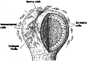
A possible model
for the initial cleft formation in the epithelium of mouse embryonic
submandibular gland. The formation of one cleft in a lobule is hypothetically
depicted. In this model the narrow cleft, like a furrow, is made by
contracting a bundle of fibrils (schematically drawn as a rope-like
structure) at the epithelial-mesenchymal interface by mesenchymal
cells. Another terminal of the mesenchymal cell mass, which takes
part in the traction, may be anchored to the lateral aspect of the
lobule or to the stalk where heavy depositiion of collagen takes place.
Although mesenchymal cells are depicted to be widely distributed,
the epithelium is sheathed in a capsule of mesenchymal cells in situ(Nakanishi
et al., J. Embryol. exp. Morph.,96, 65-77, 1986). |
|||||

|
|||||
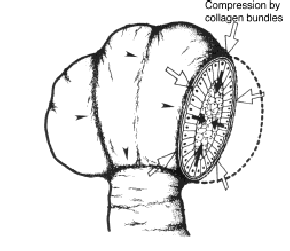
| Modified scheme of the putative action of collagen bundles at the epithelial-mesenchymal interface of submandibular gland. The depicted alignment of collagen bundles is based on the observations with scanning and transmission electron microscope and also on the three-dimensional computer reconstruction from serial paraffin sections stained with anti-collagen III antibody (Ichikawa and Nakanishi, Acta Histochem. Cytochem.,25, 623-628, 1992). One of the possible roles of collagen bundles encircling the lobule is to compress the basal side of the epithelial layer throughthe contractile activity nad flowing movement of mesenchymal cells (large arrows). Alternatively, collagen bundles send to epithelial cells a signal of invagination through the interaction with basal lamina or cell surface receptors (small arrows). Arrowheads indicate collagen bundles (Nakanishi and Ishii, BioEssays,11, 163-167, 1989). | |||||
|
上の仮説で要求される上皮の性質: Properties of the epithelial tissue required for above hypothesis: |
|||||
|
In
the 12-day and 13-day stages, desmoplakins I/II (major desmosomal
proteins) and occludin and ZO-1 (tight junctional proteins), but not
the E-cadherin/b-catenin complex, were hardly detected in the epithelial
lobules while all these molecules were present in the stalk region
and in the oral epithelium. It is of note that the E-cadherin/b-catenin
complex and the associated microfilaments were uniformly distributed
along the cell periphery in the lobules, indicating that adherens
junctions had not yet formed. In the 14-day glands, expression of
these molecules associated with cell-cell adhesion systems became
evident especially in the forming cavitated regions. These data indicate
that cell-cell adhesion systems are less developed in the early epithelial
lobules packed with cells and that there is a close relationship between
changes in tissue architecture of the epithelium and those in the
organization of cell-cell adhesion systems during the development. (Still under construction) |
|||||