|

| |||

|
||||

|
||||
| Fig. 6. High magnification view of the area indicated by the left arrow in Fig. 5, indicating that the fibre consists of many thinner fibrils. x 4030. | ||||

|
||||
|
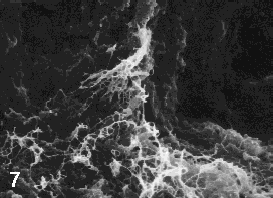
Fig.
7. Higher magnification view of the area indicated by the right arrow
in Fig. 5, showing the presence of a bundle of fibrils between two
adjacent lobules. x 2430. (Please see the following paper: Nakanishi. Y. et al., Scanning electron microscopic observation of mouse embryonic submandibular glands during initial branching: preferential localization of fibrillar structures at the mesenchymal ridges participating in cleft formation. J. Embryol. exp. Morph., 96, 65-77, 1986). |
||||